
Wright-Giemsa Stain
Intended use:
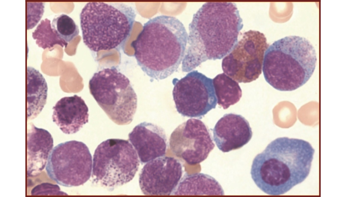
This kit is for classification of hematocytes in blood and marrow.
Principle:
Wright-Giemsa Stain, as modified from Romanowsky Stain technique, is intended for staining smears of blood and marrow.
Staining of cells involves physical adsorption and chemical affinity which allows stain to penetrate and remain wiithin cells. Becasuse each cell and its components are different in chemical composition, their affinity to acid stain (Eosin) and alkaline stain (Methylene blue) of this kit varies significantly. After staining the smear with Wright's Stain, different types of cells will appear in different colors. Consequently, one can identify cells based on their stain color, shape and other physical characteristics.
Methods:
1. Add Wright-Giemsa Solution A (about 0.5~0.8ml) to the smear and allow to cover the whole specimen. Stain
for 1 min.
2. Add Wright-Giemsa Solution B (2~3 times of Solution A) onto Solution A and mix thoroughtly with a rubber
pipette bulb. Allow to stain for 5~10mins.
3. Rinse with water gently, dry and examine the finished slide under a microscope.
Specifications:
| Contents | 6vialsx20ml/kit | 4Btlsx250ml/kit | 4Btlsx250ml/kit | 4Btlsx250ml/kit | Components |
| Wright-Giemsa A | 2x20ml | 2x250ml | 4x250ml | / |
Eosin, Methanol, Methylene Blue |
| Wright-Giemsa B | 4x20ml | 2x250ml | / | 4x250ml | Buffer |
Precaution:
1. The length of staining depends on specimen type, smear thickness, number of nucleoli, cell type and room
temperature etc. For blood smear, typically stain for 3-5mins after adding Solution B; for bone marrow
smear, stain for more than 10mins.
2. For bone marrow smear, as there are many fibrins that will clot quickly, the process of smearing must be fast.
Do not use oxalate as anticoagulant; doing so would cause the nucleolus to distort, chromatin to become
compact, cytoplasm vacuole to form and oxalate crystal to appear.
3. The staining should be performed with a sufficient volume to avoid sample drying.
4. For hematocyte stain, if the weather is cold or humid, the smear should be incubated at 37Īµ in order to
prevent cells from shrinking.
5. Cap the reagent bottle immediately after use to avoid vaporization.
6. Do not use the reagents beyond the stated expiration date. For kit storage, avoid exposure to extreme high
or low temperature and sunlight.